Synthesis and characterization of TiO2 nanowires for photovolatic and electrochemical applications
TiO2 nanostructures are promising electrodes for dye or hybrid solar cells and are utilized as stable support for electrocatalyst. Among a great variety of nanostructures, single-crystalline TiO2 nanowires exhibit particularly interesting characteristics. In this project TiO2 nanowires are synthesized via a hydrothermal approach and analyzed using electron microscopy. The main focus is on the morphology changes during annealing and core etching which lead to better performance.
Semiconducting metal oxides play a key role in photophysical and electrochemical applications, like photovoltaics, photo(electro)catalysis or water-splitting. Among these metal oxides, TiO2 is one of the most attractive electrode materials for hybrid, dye sensitized or perovskite solar cells. As these solar cells mimic the principle of photosynthesis, their performance mainly depends on charge-separation processes. Thus charge recombination and effective light harvesting highly influence the efficiency of these solar cells. Similar, this processes are also important when TiO2 is used as photocatalyst. If TiO2 is applied as support material for electrocatalyst, its chemical stability is of upmost importance.
This project focuses on the synthesis and electron microscopic investigation of highly crystalline TiO2 nanowire arrays. These one-dimensional nanostructures show a large surface area and a direct electron path to the substrate and thus are promising for integration in high efficiency solar or (photo)electrochemical cells. The TiO2 nanowires are further modified by annealing in different atmospheres and by etching with the aim to change their conductivity and to enhance their surface area. Homogeneous deposition of nanometer-sized metal catalyst is also carried out and the electrocatalytic property and stability of the nanostructures is tested.
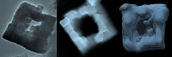
Advanced transmission electron microscopy (TEM) techniques are used to analyze these nanowires before and after the heat treatments and electrochemical reactions. To see structural changes in between, in-situ heating experiments in the TEM and electron energy loss spectroscopy (EELS) analyses are performed. TEM tomography is also used to reconstruct the 3D morphology of the nanowire.

(a) TiO2 nanowire after core etching, (b) 3D reconstruction of hollow TiO2 nanowire, and (c) metal nanoparticles grown on hollow TiO2 nanowires.

