The TOMO Project – Integrating a Fully Functional Atom Probe in an Aberration Corrected TEM
Joachim Mayer1,2* , Juri Barthel1 , Ashok Vayyala1 , Rafal Dunin-Borkowski1 , Maarten Bischoff3 , Hugo van Leeuwen3 , Stephan Kujawa3 , Joe Bunton4 , Dan Lenz4 , Thomas F. Kelly5
1. Ernst Ruska-Centre, Forschungszentrum Jülich GmbH, Jülich, Germany
2. Central Facility for Electron Microscopy, RWTH Aachen University, Aachen, Germany
3. Thermo Fisher Scientific, Eindhoven, The Netherlands
4. CAMECA Instruments, Inc., Madison, WI, United States
5. Steam Instruments, Inc., Madison, WI, United States.
* Corresponding author: Prof. Joachim Mayer
In the present contribution, we want to introduce the TOMO microscope which will be based on a
combination of two well-known but fundamentally different materials analysis techniques in one device:
a state-of-the-art atom probe will be integrated into a high-performance TEM. This plan represents a
revolutionary step towards atomic-precision analysis and has not previously been realized. The first
important advantage results from the almost complete complementarity of the techniques, which we will
be able to apply to the same object simultaneously [1-4].
In a correlative approach, the segregation of a specific element to a defect or any other microstructural
feature in a given matrix phase can most sensitively be analysed with the atom probe technique.
However, the atom probe cannot deliver atomically resolved information on the atomistic structure and
the local bonding situation, which in a complimentary way can be given by HRTEM/STEM and EELS.
Although experimental workflows exist in which an APT needle is first characterised by TEM, reliable
results can only be obtained for the thinnest part of the needle, but not for defects which will only be
exposed after further ablation of the needle. On the other hand, the standard method of reconstruction of
the APT atomic positions assumes that the apex shape of the needle is hemispherical. If the apex is not
spherical, then errors in atom positioning result. In the presence of dislocations, twin boundaries,
stacking faults, grain boundaries or multiple phases in the sample, grooving occurs which leads to an
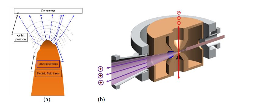
alteration of the trajectories and thus to artefacts in the final APT tomograms, see also Figure 1.
The TOMO instrument, which will be installed at the Ernst Ruska-Centre in 2024, will benefit from the
almost complete complementarity of the techniques, which can be applied to the same object
simultaneously. While the TEM offers high spatial resolution from a small sample area with relatively
modest elemental sensitivity (approximately 1 %), the atom probe has lower spatial resolution but
unrivalled elemental (isotopic) sensitivity in the part-per-million (ppm) range. The expected synergistic
gain obtained by combining the technologies results from the fact that it will be possible to observe the
shape of the tip apex during an experiment and thus correct atom trajectories for greater spatial
precision. Furthermore, the true electric extraction field can then either be calculated from the image of
the tip shape or it can be measured directly in the electron microscope by holographic techniques.
For the first time, the TOMO instrument will permit the determination of the types and locations of
millions of atoms in a volume of several hundred thousand cubic nm, down to each individual lattice
position, in one measurement. The three-dimensional distributions of functional elements can be studied
to almost the single-atom level, while linking it to atomic structures with picometre precision and
electronic structures with sub-eV resolution. The structures and defects of relevant material systems like
nanoelectronic components, compound solar cells or catalytically active nanoparticles, for example, can
be imaged and analysed in parallel. In addition, the segregation of light elements such as H, Li or O to
active defects and interfaces can be examined, leading to novel insights into a wide variety of structural
materials, energy storage systems and functional elements [5].

how the electric field line geometry influences the ion trajectories and registration positions on the two-
dimensional detector. (b) Schematic drawing of the integration of the APT needle in the centre of the
pole piece lens of the TOMO-TEM. UHV conditions and temperatures below 35 K will be ensured by a
completely new column and stage design.

[1] Michael K. Miller, Thomas F. Kelly, Krishna Rajan, and Simon P. Ringer, Materials Today, April
2012, vol. 15, no. 4, pp. 158-165.
[2] Thomas F. Kelly, Michael K. Miller, Krishna Rajan, and Simon P. Ringer, Microscopy and
Microanalysis, Invited Review, vol. 19 (2013) pp. 652 – 664.
[3] Thomas F. Kelly, Microscopy and Microanalysis, vol. 23 (2017) pp. 34-45. doi:10.1017/S1431927617000125
[4] Thomas F. Kelly, Brian P. Gorman, and Simon P. Ringer, “Atomic-Scale Analytical Tomography: Concepts and Implications,” (Cambridge University Press 2022).
[5] The authors acknowledge funding from the German Ministry of Education and Research (BMBF) in the framework of the National Research Infrastructure Roadmap.

