© G. Geelen, Max-Planck-Institut für Eisenforschung GmbH

Equipment and Facilities

The Department Structure and Nano- / Micromechanics of Materials employs dedicated techniques and advanced facilities, which are organized in five laboratories.

Advanced Transmission Electron Microscopy

The SN department utilizes the following transmission electron microscopy (TEM) techniques:

  • Conventional TEM imaging and selected area electron diffraction
  • Electron energy loss spectroscopy (EELS) and energy-filtered TEM (EFTEM) imaging
  • Aberration-corrected scanning TEM (STEM) in combination with energy dispersive X-ray spectroscopy (EDS) and EELS
  • In-situ heating and in-situ nanoindentation
  • In-situ liquid flow cell holder

Micro- and nanomechanics in situ

Probing material properties at the micron/nano scale requires dedicated machines and setups for sample manufacture, sample testing, and in situ observation as well as post mortem defect analysis. The following capabilities are available to produce and deform micron/nano sized samples:

Focussed ion beam milling:

  • Ga-focussed ion beam (FIB) system operated between 5 keV to 30 keV
  • Nano patterning and visualisation engine with full beam control access to shape complex patterns
  • Integrated electron backscatter diffraction (EBSD) for accurate determination of crystal orientations
Miniaturised mechanical testing:
  • Commercial and in-house fabricated nanoindentation systems for operation inside scanning electron microscopes (SEMs) and at synchrotron beamlines
  • Custom-modified in situ micro/nano mechanical testing system capable of accessing a wide range of strain rates and temperatures
  • Nanoindenter holder for the transmission electron microscope (TEM)

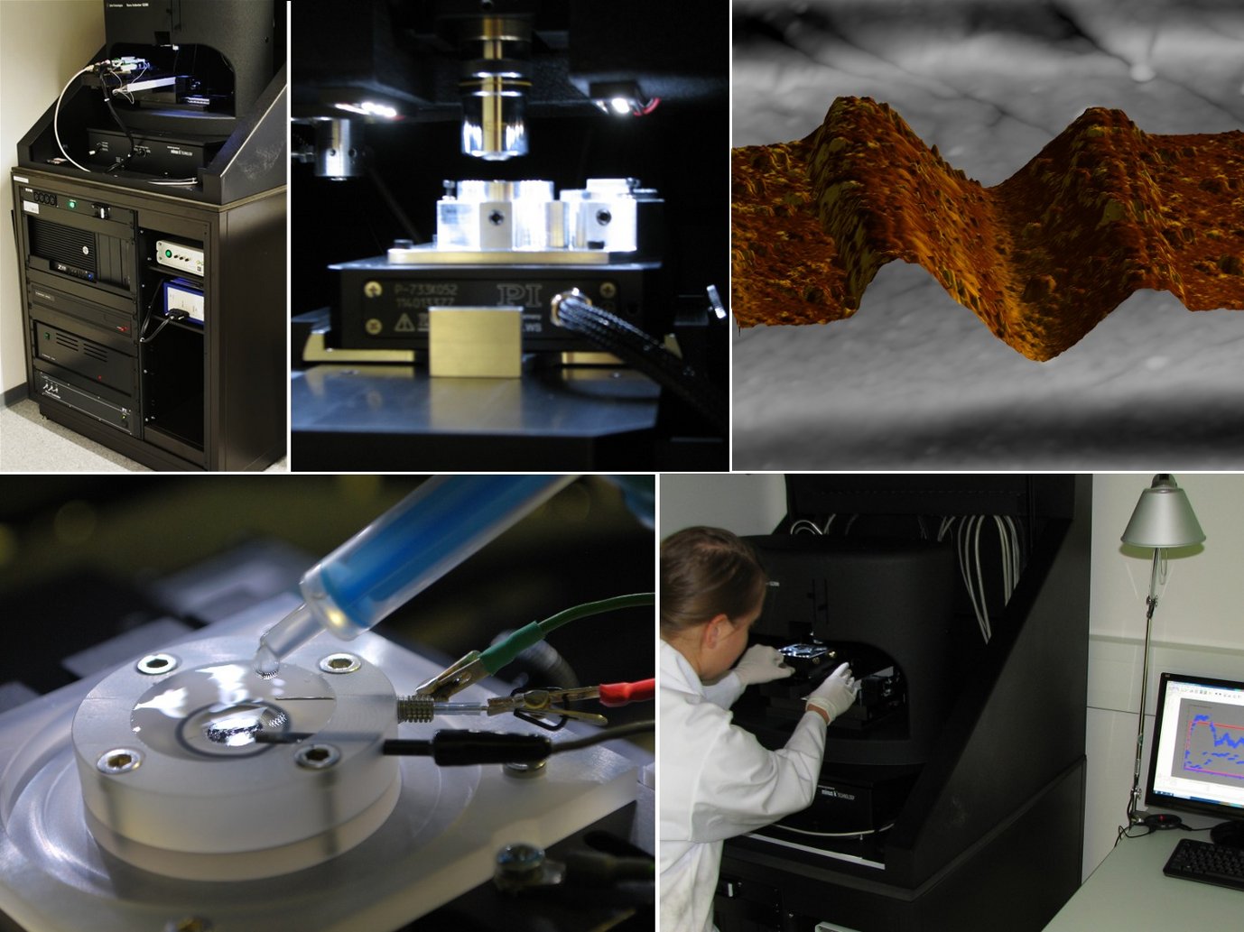
Nanoindentation and Nanotribology

Nanoindentation and –tribology rely on measuring nanometer displacements inside a KLA G200 system with the following options::

  • XP and DCM II heads for nanoindentation measurements with a maximum normal force of 0.5 N and displacement resolution <1 nm
  • High positional accuracy
  • Lateral force measurements
  • Fast indentation testing for hardness and modulus mapping
  • Front-side three-electrode electrochemical cell and high accuracy potentiostat, for hydrogen charging and corrosion testing during mechanical testing
  • Back-side three-electrode electrochemical cell for hydrogen charging during mechanical testing

Typical experiments:

  • Young’s modulus and hardness measurements (standard and mapping)
  • Materials: bulk, thin films on substrate, micrometer sized phases in bulk
  • Topographic scanning of surface
  • Hydrogen embrittlement
  • Friction and scratch experiments of metals under different environments

Thermal Analysis and Electron Probe Microanalysis

For thermal analysis the following techniques are available:

  • Differential thermal analysis (DTA) between 140 K and 2000 K
  • Differential scanning calorimetry (DSC)

Electron probe microanalysis (EPMA) by wavelength-dispersive X-ray spectrometry (WDS) focusses on

  • Chemical compositions of individual phases
  • Concentration profiles, segregations, element mapping

X-Ray Diffraction

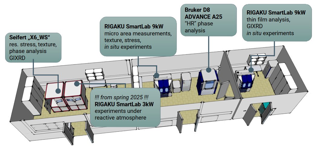
The X-ray laboratory is equipped with four different diffractometers. In 2012 the complete laboratory infrastructure was recently reorganized in a single x-ray lab. The laboratory equipment is continuously updated. Two new diffractometers were installed in 2019 and a new device was installed 2023. An additional fifth diffractometer for XRD experiments under reactive atmosphere will be installed in spring 2025.

A wide range of X- ray techniques are possible:

  • phase analysis (qualitative and quantitative)
  • residual stress analysis
  • texture analysis
  • characterization of thin layers
  • XRD measurements during load or heat
  • XRD measurements under reactive atmosphere (from spring 2025)

The x-ray laboratory could handle different sample geometries:

  • powder samples
  • constructive parts (maximum weight 10kg)
  • small solid samples (minimum beam diameter 0.7mm)
  • thin layers
  • single crystals
  • weld samples
Go to Editor View