Orientation (Imaging) Microscopy - ORM/OIM
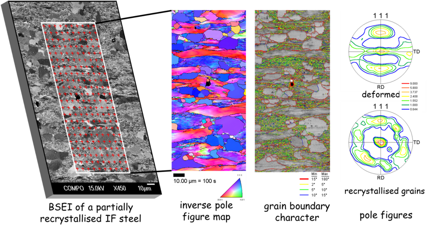
A (scanning) electron microscopy technique which measures crystal orientations on a regular grid by electron diffraction. The so-determined data are used to produce orientation or phase maps of the scanned area on the sample.

We refer here exclusively to the technique based on electron backscatter diffraction (->EBSD). When the electron beam is scanned over a crystalline sample, an EBSD pattern is obtained for every scan grid position. Using software, the orientation, crystallographic phase and lattice perfection can be obtained in a fully automated manner from each of the patterns. In this way a large data set is produced that quantitatively represents the scanned area of the sample. These data can subsequently be used to reconstruct the microstructure of this area, for example by assigning similar colours to points with similar crystal orientation or similar phases. In this way orientation maps are obtained. By determining the change of orientation from position to position, misorientation maps can be produced as well. Besides a large variety of different maps it is also possible to construct statistical data sets from the sample, for example orientation distributions (textures) displayed, e.g., in form of pole figures of different constituents of the mapped area (here: deformed and recrystallized grains).
Some characteristic data: lateral resolution: 20 to 100 nm (depending on the sample material); measurement rate currently up to hundreds of patterns per second; Investigated areas: few µm² to few cm². Angular resolution: 0.5° down to 0.01° with special techniques (->XR EBSD). The technique can also be extended into a 3D technique (-> grain boundaries).
