3D EBSD-based Orientation Microscopy (3D EBSD)
EBSD-based orientation microscopy is by nature a 2-dimensional technique, revealing the microstructure existing close to the surface of the sample. By combining 2D-ORM with successive serial sectioning a 3D technique is developed. 3D microstructures are reconstructed from the individual sections by dedicated in-house software.

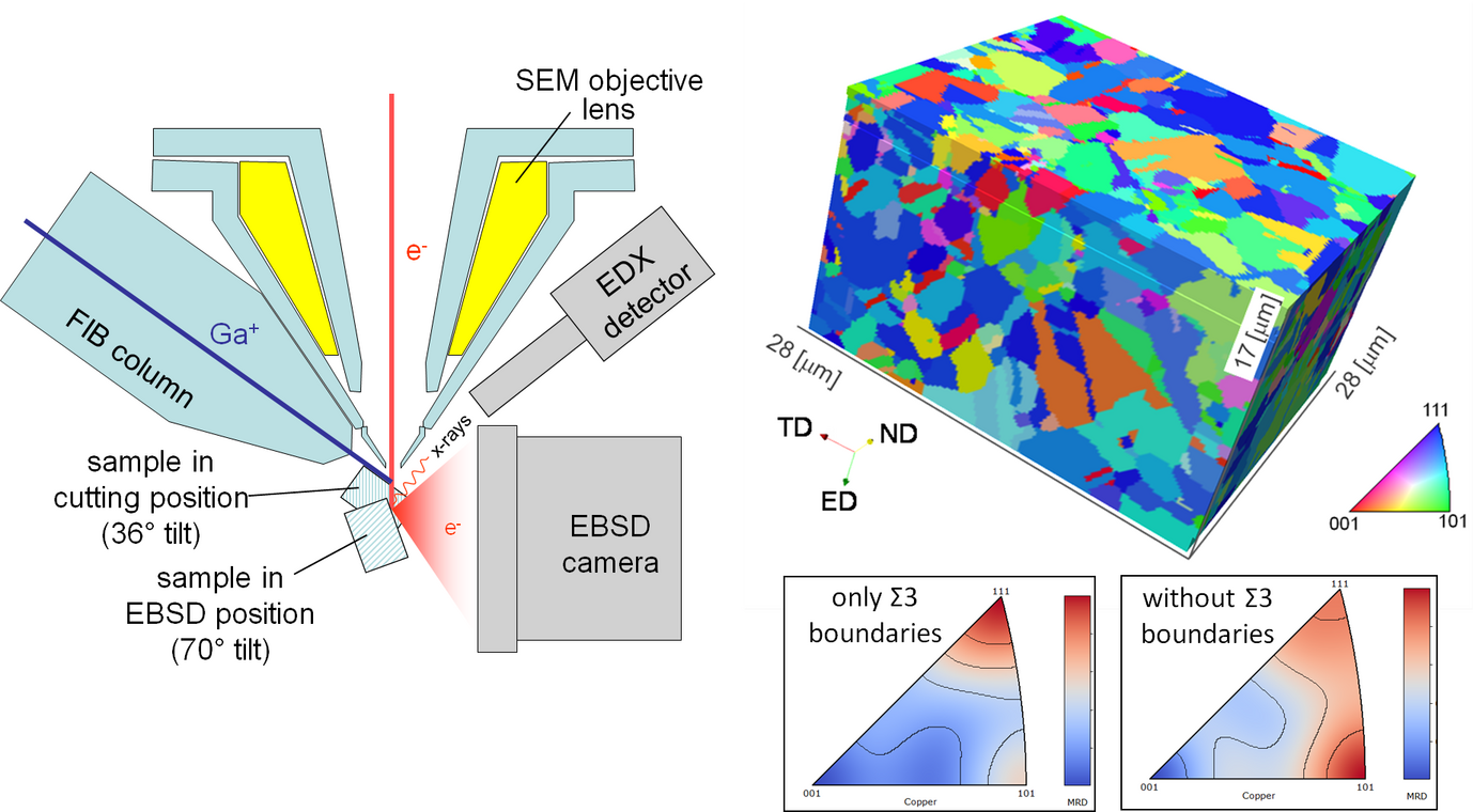
Right: 3D ORM of a severely deformed and recrystallized Cu-Zr alloy. Colours correspond to the inverse pole figure position of the sample normal direction.
Bottom: Inverse pole figures of the grain boundary normal vector distributions for Σ3-misoriented grains (twins) and for all other grain-pairs.
Suitable serial sectioning methods are milling with a focused ion beam (FIB) in gracing incidence or mechanical polishing. We have, so far, developed a fully automated FIB-based method. It is installed on a Zeiss XB 1540 FIB-SEM cross beam instrument. Using a stage tilt the sample is alternately moved to milling in gracing incidence and to EBSD-based orientation mapping. The correct position is checked by detection of a fiducial marker with an image recognition system. In this way a spatial resolution of 50 nm can be reached. The technique captivates by accurate slice thickness and parallelism, as well as relatively easy automation. We have measured successfully different sorts of steels (both austenitic and ferritic), Mg-, Al-, Cu, Ti-, and Ni-alloys, superalloys, and Cd-Te structures. Disadvantageous is, however, the small total volume measurable by FIB-based serial sectioning, which is limited to a maximum of about 50 × 50 × 50 µm³ (usually on 20 × 20 × 20 µm³), the relatively low slicing rate, and FIB-induced lattice defects and artefacts. The latter may make measurements by EBSD completely impossible. Artefacts have been observed on many materials with non-metallic bonding (intermetallics, carbides) and on metastable structures (metastable austenite). A serial sectioning method by mechanical polishing appears, therefore, desirable.
3D-OMI is particularly important for the determination of the comprehensive characterization of grain boundaries with all 5 rotational degrees of freedom being revealed. Furthermore, 3D-OMI may help understanding complexly shaped microstructures (e.g. interconnected phases in eutectic structures, cracks, serrated boundaries). Finally, 3D-OMI may be used to determine the density of geometrically necessary dislocations (GNDs). In contrast, 3D OMI is, in most cases, not a suitable technique to measure grain sizes, grain morphologies, phase fractions and textures, because of the low statistical significance of the data.
