Mapping Hydrogen in TWIP Steels
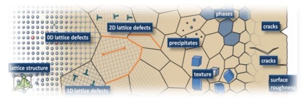
Hydrogen embrittlement of austenitic steels is of high interest because of the potential use of these materials in hydrogen-energy related infrastructures. In order to elucidate the associated hydrogen embrittlement mechanisms, the mapping of heterogeneities in strain, damage (crack/void), and hydrogen and their relation to the underlying microstructures is a key assignment in this field.
In the last years several promising novel approaches have been reported for the localized resolved and sensitive detection of hydrogen.
One is a direct electrochemical detection via a capillary cell, developed by Suter et al. which, however, does not provide the resolution required here. Another approach is the use of Kelvin probe techniques, which allow detection of hydrogen in a material by means of the change ofwork function caused by the hydrogen entering the oxide at the surface. Studies at quite high resolution have been carried out with Scanning Kelvin Probe ForceMicroscopy (SKPFM), where diffusion profiles of hydrogen have been mapped successfully at relatively high resolution at cross section of samples after hydrogen charging.
However, a direct quantification is not possible by this method, due to the complex dependence of the work function of oxide on different defect states in the oxides. For the same reason this approach is not suitable to provide reliable information on hydrogen at different features of the microstructure, because different oxides show a different dependence on hydrogen. A new approach by applying a thin palladium layer eliminates this problem.