O‘Hydrogen where art thou?
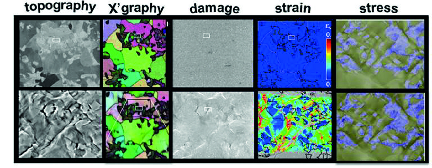
One of the experimental goals of the Adaptive Structural Materials group is to understand the hydrogen embrittlement micro-mechanisms in advanced high strength alloys (AHSA). The challenge enroute is immense, since most novel AHSA have complex micro-composite structures consisting of multiple phases of high mechanical contrast. To simultaneously deliver high strength and toughness, these phases often differ in crystal structure, mechanical-stability, defect density, active deformation or cracking mechanisms etc. with increasing deformation each phase evolves differently, essentially resulting in a different micro-composite response at different deformation levels. In presence of hydrogen the picture gets even complexer: First of all, different phases have different solubility of hydrogen. Second, hydrogen diffusion kinetics differ in different phases. Third, hydrogen may cause different embrittlement mechanisms in different phases. This complex process clearly can-not be fully understood by traditional ‚post-mortem‘ approaches. We approach this as a multi-field mapping problem. By employing various tools and techniques the evolution of microstructure, micro-strains, micro-stresses and micro-damage in such complex AHSA, can be mapped, in-situ, at high resolution (Fig. 1).

Fig. 1: Mapping of deformation induced evolution of local microstructure, strain, stress, damage fields by (from left to right) SE imaging, EBSD, Inlens SE imaging, microscopic- DIC, crystal plasticity simulations. Shown data is of a dual phase steel.
To map the deformation-induced evolution of microstructure, high resolution electron backscatter diffraction and electron channelling contrast imaging techniques are used. Micro-strains are tracked by microscopic digital image correlation, using in-lens se images and fine SiO2 particles. Micro-stresses are experimentally difficult to measure, thus we rely on crystal plasticity simulations. Micro-damage incidents are characterized by inlens se imaging and post-mortem serial sectioning & imaging.

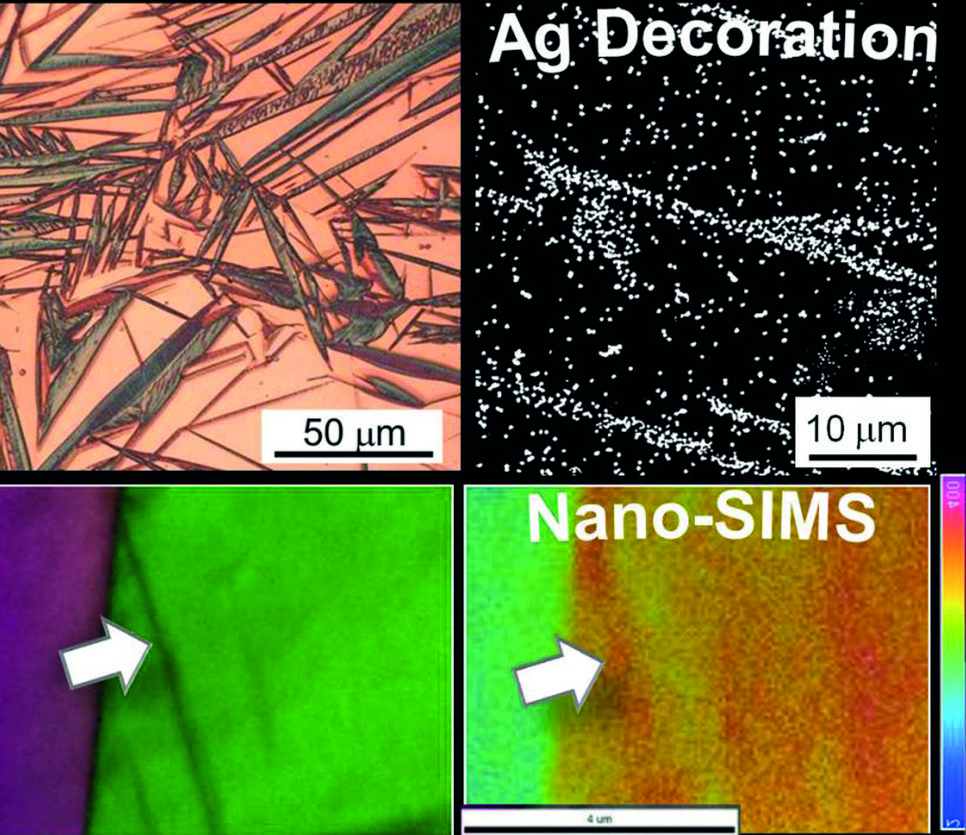
Fig. 2: Hydrogen mapping by: Ag decoration (above) in martensitic-austenitic steel (collaboration with the Kyushu University); Nano-SIMS (below) in a TWIP steel (collaboration with RWTH Aachen and LIST). In the former higher hydrogen content is observed in martensite plates, while in the latter, in deformation-induced mechanical twins.
What about hydrogen? In collaboration with research partners inhouse (i.e. the group “Corrosion” of Dr. Michael Rohwerder), at the Kyushu University, and at The Luxemburg Institute of Science and Technology (LIST), various techniques are explored, each of which with different advantages and limitations. These include scanning Kelvin probe force microscopy (SKPFM), secondary ion mass spectroscopy (SIMs or Nano-SIMs), Ag decoration, all assisted by thermal desorption spectroscopy (TDS). some examples are shown in Fig. 2.
The group “Adaptive Structural Materials” has so far applied the approach to TRIP-Maraging steels, martensitic steels, dual phase martensitic-ferritic steels, and martensitic-austenitic steels. While the incorporation of the hydrogen mapping techniques to in-situ analysis during deformation remains challenging, the overall approach still provides a unique overview of all the inter-linked micro-processes taking place during deformation. The realm of data that is produced in this manner reveals great insight on how novel alloys can be developed for better hydrogen resistance.
Author: Cem Tasan

