Artificial Intelligence in Materials Science
The amount of data produced by detectors has increased explosively. Automated tools to process and analyze the data are necessary. Artificial intelligence (AI), in particular machine learning (ML), has been exploited by an increasing number of disciplines to automate complex problem-solving tasks. Progress in Ml has led to decision rules that can in some cases be automatically derived by specific algorithms. One of the successful applications of ML in materials science lies in developing fully automated algorithms for analyzing high-throughput experiments.
Field ion microscopy (FIM) is a high electric field technique, which enables the imaging of surfaces with atomic resolution. When exposing the tip not only to the minimum field strength required for ionizing the imaging gas but also for evaporating the tip atoms themselves continually, the method is rendered depth sensitive (3D-FIM). This means that the specimen can be investigated tomographically along the tip’s longitudinal axis. 3D-FIM is capable of producing large and accurate tomographic datasets containing information on sequential atomic positions, but these large datasets lead to a new tremendous challenge of how to manage the data.
Presently, there is a lack of efficient data handling algorithms to extract pertinent information from these data-sets in an (a) automated; (b) fast; (c) user-independent; and (d) error quantified manner. For instance, characterization of a volume of 0.001 μm3 (a typical 3D-FIM sample size) produces in the range of 2 × 10⁵ images. Hence, there is a great need for efficient al-gorithms and data mining routines. To this end, we recently proposed a new method to extract atomic positions from 3D-FIM datasets, using various image processing and machine learning algorithms [1, 2].

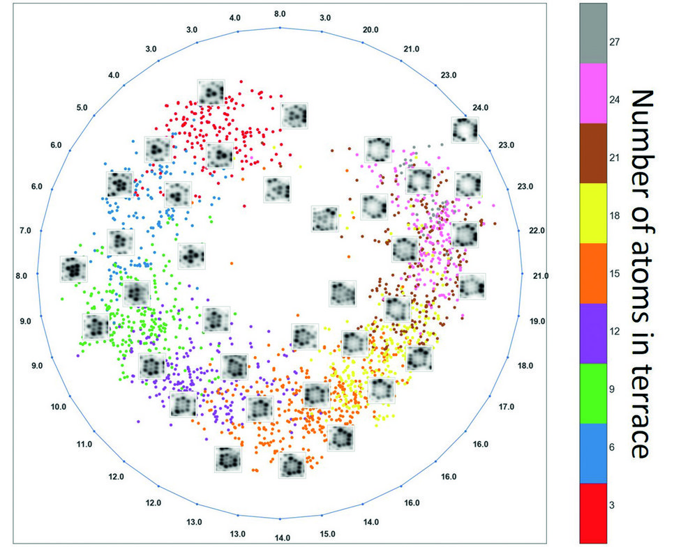
Fig. 1: Machine learning (Isomap) on the 3D-FIM dataset. Each point represents one picture in the reduced dimensional manifold, and its colour indicates the number of atoms in the first terrace of the corresponding picture. The numbers on the outer circle represent the averages over the number of atoms in the first terrace of pictures.
Applying machine learning to the 3D-FIM images (> 21000 dimensions) and projecting the data to a low dimensional subspace helped us to understand the latent structure in the data. The exemplary result in Fig. 1 reveals a cyclic behaviour in the dataset, which is a consequence of the field evaporation behaviour. Field evaporation typically occurs layer-by-layer and proceeds from atoms sitting at the ledge of a terrace towards the centre. When a terrace field evaporates the atoms sitting on the ledge disappear, decreasing the number of atoms on the terrace as the evaporation proceeds. By evaporating atoms from the surface of the sample during FIM, the first terrace area decreases until it evaporates completely. The number of cycles is a measure of the number of atomic terraces evaporated during the 3D-FIM.
With the use of such advanced algorithms for data extraction, we hope not only to improve the accuracy of the data extraction from 3D-FIM but also to identify and characterize various material defects. Currently, the physics of image formation in FIM are still not completely understood, but it is our firm belief that ML can be a powerful tool in this direction.
References:
Authors: Gh. A. Nematollahi, B. Grabowski
