Atomic Electron Tomography Using Coherent and Incoherent Imaging in (Scanning) Transmission Electron Microscopy
Atomic Electron Tomography Using Coherent and Incoherent Imaging in (Scanning) Transmission Electron Microscopy
- Date: Oct 18, 2018
- Time: 10:00 AM - 11:00 AM (Local Time Germany)
- Speaker: Dr. Colin Ophus
- NCEM, Molecular Foundry, Lawrence Berkeley National Laboratory
- Location: Max-Planck-Institut für Eisenforschung GmbH
- Room: Seminar Room 1
- Host: on invitation of Dr. Christian Liebscher and Prof. Gerhard Dehm
- Contact: stein@mpie.de

The past decade of development for transmission electron microscopy (TEM) and scanning TEM (STEM) have been enormously successful. In structural biology, cryo-TEM methods can solve the structure of proteins with sub-nanometer resolution. In materials science, hardware aberration correction has enabled routine atomic resolution imaging in two dimensions, as well as allowing 3D tomography that can identify the 3D position and species of every atom in a nanoscale sample. This technique, called atomic electron tomography (AET), has been applied using high angle annular dark field (HAADF)-STEM, which is a high dose imaging method that not sensitive to weakly scattering elements such as a carbon or oxygen. By contrast, cryo-TEM is used to reconstruct the 3D structure of biological structures which are composed of only weakly-scattering elements. This is accomplished by averaging hundreds or thousands of images of identical or near-identical samples, which is only possible because of biological replication of proteins. Thus, in materials science there is a need to solve the structure of unique, heterogeneous samples composed of weakly-scattering or beam-sensitive samples.
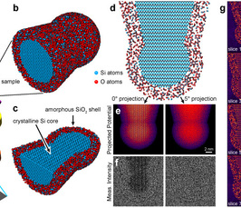
In this talk, I will show our AET results for FePt nanoparticles, including multiple time steps of the same particle after annealing steps. These results show atomic-scale nucleation and growth for the first time in 3D. I will also present a practical method for reconstructing the electrostatic potential from a tomographic tilt series of HRTEM phase contrast measurements, including multiple scattering and strong phase shifts. I test the method using multislice simulations of a sample consisting of a crystalline silicon core, surrounded by an amorphous silicon dioxide shell. These simulations show that our method is robust to a restricted tilt angle range, uses much lower electron doses than existing AET studies, and can be used for a wide range of experimental parameters. The experimental geometry, a simulated test sample, projected images and reconstruction are shown in Figure 1, adapted from arXiv:1807.03886. I will also show our current progress in reconstructing experimental phase contrast HRTEM tilt series datasets at atomic resolution.